Three Dimensional Display Systems for Surgery in Ophthalmology

Microsurgery has witnessed a significant increase in the last two decades due to new innovations. However, the traditional binocular microscope used in microsurgery can lead to musculoskeletal fatigue and injuries, reducing the surgeon's longevity. Ophthalmologists experience neck, upper body, or lower back symptoms due to the traditional microsurgery posture.

Three-dimensional (3D) display systems were first developed for aircraft and military use and were later brought to the operating theater. Live 3D display systems have been widely used in medicine, particularly after the creation of the TrueVision 3D Visualization System for Microsurgery. Conventional 3D systems are categorized as either active or passive systems.

In active systems, high-speed consecutive images for the right and left eyes are shown alternatively, while a special pair of electronic glasses actively suppresses the image in the other eye. In passive 3D systems, the three-dimensional image is acquired by mixing two images horizontally and then passively separating them into polarized 3D glasses.

In ophthalmology, heads-up surgery describes microsurgical procedures performed by viewing the microscopic image on a panel display sent from a 3D camera instead of looking at the eyepieces of the microscope. This technique eliminates the constraints imposed by the standard binocular microscope and provides greater degrees of freedom to operate in a more neutral, physiologic position without affecting the image quality or technical difficulty. It is also potentially an aide to surgical teaching since multiple people in the same room can share the high-resolution view of the surgeon.

Cataract and anterior segment surgery in human eyes using heads-up surgery were first reported by Weinstock in 2010. He reported excellent outcomes in both groups, with minimal procedure time difference between groups. Interestingly, the rate of unplanned vitrectomy was three times higher in the standard microscope group compared with the TrueVision group.

The TrueVision 3D Surgical System is a camera unit that attaches to standard surgical microscopes, sending stereoscopic images and video to a 3D, high-definition (HD), large-screen monitor positioned a few feet from the surgeon, providing visualization in real-time. The US Food and Drug Administration (FDA) has granted clearance for the TrueVision Refractive Cataract Toolset, an application that provides 3D graphical overlays for image-guided cataract surgery. 

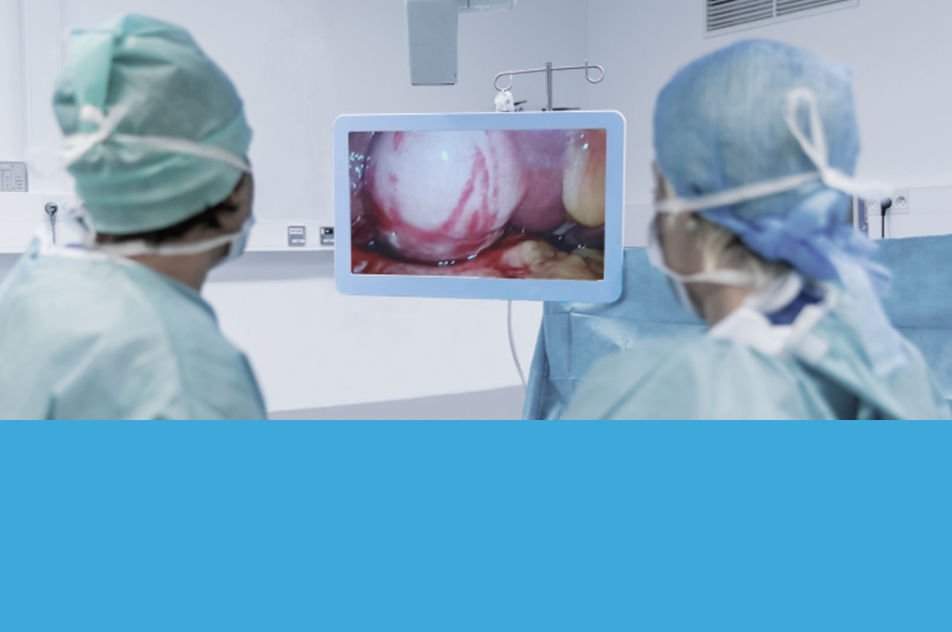
Other anterior segment surgeries have been performed using the heads-up system, including amniotic membrane transplantation and corneal surgery. Vitreoretinal procedures have also been performed using the heads-up surgery, which was introduced by Eckardt and Paulo. They conducted a study to assess whether vitreoretinal surgery could be performed using the heads-up display system with a 3D, high dynamic range surgical camera, a HD LCD display which requires the use of passive 3D polarized glasses.

The use of head-mounted display systems is a novel emerging concept in ophthalmology. The head-mounted system differs from conventional 3D systems by showing two simultaneous images, one for each eye, avoiding the ghosting image effect caused by cross-talk in active 3D systems. The wide horizontal viewing angle enables a more natural visual experience. The use of this technology in Ophthalmology has been widely reported, including pars plana vitrectomy, alone and combined with phacoemulsification/intraocular lens (IOL) implantation.

In conclusion, 3D display systems have shown promising results in ophthalmology, with heads-up surgery using 3D display screens increasingly accepted due to their advantages in terms of ergonomics and surgical teaching/coordination. The use of head-mounted display systems is an innovative concept in ophthalmology that may prove to be an excellent tool for live surgery teaching and training in the future.